So I've had the second bout of knee surgery and these are the pictures of the inside of my knee...
| 1 | |
2 |
| 3 | 4 | |
| 5 | 6 | |
| 7 | 8 |
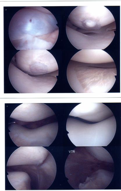
1. This is a shot through my knee which shows the membrane I shouldn't have. It's a pre-birth relic, they should get reabsorbed and usually do. About 10% of the population have remnants, but it's extremely rare to have a complete septum. The hole in it was made by the surgeon while he was investigating.
2. This is the site of the previous repair. The lesion in the cartilage was drilled out and the underlying bone was fractured. The regrown cartilage has overgrown a bit.
3. This is after it being flattened out. The colour difference between the regrown cartilage and the original is obvious.
4. Surgeon also found this; it's apparently just calcium deposits in the cartilage, which is perfectly normal, he's just never seen them in that sort of patterning before.
5. This is my kneecap after the removal of the septum, nicely positioned over the groove. in my femour.
6. This is the position before -- it doesn't quite sit in the groove.
7. This is one of the outer sides of my knee, which looks ok
8. This is the other side, which doesn't look so good. He removed the torn cartilage -- it's in a non-weightbearing bit so it shouldn't bother me.